Foot & Ankle Treatments
Ankle Arthroscopy
Arthroscopy is a surgical procedure during which the internal structure of a joint is examined for diagnosis and treatment of problems inside the joint. Ankle Arthroscopy includes the diagnosis and treatment of ankle conditions. In arthroscopic examination, a small incision is made in the patient's skin through which pencil-sized instruments that have a small lens and lighting system (arthroscope) are passed. Arthroscope magnifies and illuminates the structures of the joint with the light that is transmitted through fibre optics. It is attached to a television camera and the interior of the joint is seen on the television monitor.
Arthroscopic examination of ankle joint is helpful in diagnosis and treatment of the following conditions:
- Inflammation: Synovitis, the inflammation of the lining of the ankle joint
- Acute or chronic injury
- Osteoarthritis: A type of arthritis caused by cartilage loss in a joint
During arthroscopic ankle surgery, either a general or local anaesthesia will be given depending on the condition. A small incision of the size of a buttonhole is made through which the arthroscope is inserted. Other accessory incisions will be made through which specially designed instruments are inserted. After the procedure is completed arthroscope is removed and incisions are closed. You may be instructed about the incision care, activities to be avoided and exercises to be performed for faster recovery.
Some of the conditions treated by ankle arthroscopy include:
- Achilles tendon rupture
- Bunion Surgery
- Sports injuries
- Toe deformities
Some of the possible complications after arthroscopy include infection, phlebitis (clotting of blood in vein), excessive swelling, bleeding, blood vessel or nerve damage and instrument breakage.
Recovery
It may take several weeks for the puncture wounds to heal and the joint to recover completely. A rehabilitation program may be advised for a speedy recovery of normal joint function. Your child can resume normal activities and go back to school within a few days.
Bunion Surgery

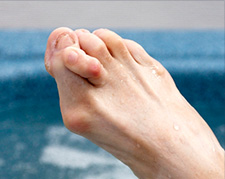
Bunion is a foot deformity that changes the shape of the foot causing the big toe to turn inward, towards the second toe leading to pain and inflammation. A bunion is caused by incorrect footwear, joint damage, arthritis, and genetic disposition. Some of the commonly observed symptoms are pain, inflammation, bony bump on the side of the foot.
Bunion may be corrected with conservative treatment or surgery. If the conservative treatment does not treat the bunion pain, then your surgeon may recommend bunionectomy, surgical removal of a bunion. The main goal of the surgery is to remove painful deformities, restore normal bone alignment, and prevent recurrence.
This surgery involves shaving the inflamed tissue around the big toe joint using a shaving drill. A part of the bone in big toe is cut to straighten the toe (osteotomy). Ligaments are tightened and adjusted in a proper direction. Screws and pins are used to fuse the bones together. After the surgery, your surgeon may instruct you to wear crutches to prevent weight bearing. Some of the complications of the surgery include infection, stiffness of the toe, loss of blood supply to the toe, and over-correction. Over-correction leads to turning of the big toe outward (Hallux varus). However, these complications can be corrected.
Flatfoot Reconstruction
Foot reconstruction is a surgery performed to correct the structures of the foot and restore the natural functionality of the foot that has been lost due to injury or illness.
Flat foot or pes planus is a condition in which the foot does not have a normal arch when standing.
Flat Foot Reconstruction:
The primary objectives of flat foot reconstruction are reduction of pain and restoration of function and appearance. This can greatly benefit patients' medical and aesthetic needs. The surgery to be performed depends on several factors such as the age of the individual, severity and duration of the symptoms.
It is often recommended when conservative treatments fail to resolve the symptoms.
The traditional method of treating flat foot is replaced by a minimally invasive technique (arthroscopy) which can be performed on an outpatient basis.
This procedure is usually performed under general anaesthesia. Several tiny incisions are made by your surgeon to insert an arthroscope and miniature surgical instruments into the joint. The camera attached to the arthroscope displays the internal structures on a monitor and your surgeon uses these pictures to evaluate the joint and direct the small surgical instruments either to repair or remove the damaged bone or tendon depending upon the extent of injury.
At the end of the procedure, the surgical incisions are closed by sutures or protected with skin tapes and a soft dressing pad is applied. Depending upon the surgery, your surgeon will place a cast or a splint to prevent movement of the foot until it regains normal functioning capacity.
Some of the advantages of arthroscopic surgery include:
- Minimal trauma to the surrounding structures
- Shorter recovery time with less post-surgical complications
- Greater range of motion with less post-operative pain
- Decreased muscle atrophy
After the surgery
Following are the post-surgical guidelines to be followed after reconstruction:
- Make sure you get adequate rest. Avoid using the affected foot for a few weeks.
- Take medications to help alleviate pain and inflammation as prescribed by your doctor.
- Apply ice bags over a towel to the affected area for about 15-20 minutes to reduce post-operative pain and swelling.
- Compression dressings (bandage) are used to support the foot to reduce swelling. Take care not to wrap too tightly which could constrict the blood vessels.
- Keep the foot elevated at or above the level of your heart. This helps minimize swelling and discomfort.
- A wheelchair might be required for a few days in more severe cases.
- Start rehabilitation (physical therapy) as recommended by your surgeon to improve range of motion.
- Crutches or a walker may be used to maintain balance or stability while walking. You should begin appropriate exercises to stretch and strengthen the muscles.
- Cover the splint while showering to keep it clean and dry.
- Return to sports once the foot has regained normal strength and function with your surgeon's approval.
The outcome of flat foot reconstruction surgery is greatly improved when you, your surgeon, and the physical therapist work together as a team.


